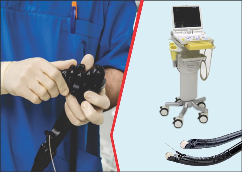
Endoscopic Ultrasound (EUS)
Endoscopic ultrasound (EUS) combines endoscopy and ultrasound to create images of the digestive tract and its surrounding organs and tissues. Endoscopy uses a long, thin tube, inserted into either the patient’s mouth or rectum that contains a camera (endoscope) to see inside the body. Ultrasound uses sound waves to create images of the inside of the body. With these two technologies, your doctor can create more detailed images of the digestive tract, Endoscopic ultrasound is a minimally invasive, outpatient procedure.
The latest endoscopic technology at Shifa International Hospital, Islamabad provides the operators with a high definition image capture of digestive tract. The procedure is performed by our internationally trained & experienced interventional gastroenterologist assisted by a dedicated team of technicians and support staff working tirelessly to provide the best healthcare services to our valued patients. An integrated reporting system is set in place to support the intervention ensuring the highest standards of care and safety
Or Dial 051 846 4646 from your Smartphone.
The state of the art Endoscopic Ultrasound (EUS) Facility at Shifa International Hospital, Islamabad is among few centers in the region offering vast range of diagnostic and therapeutic endoscopic interventions. Our doctors are extensively trained in performing endoscopic procedures for common to rare diseases and conditions.
Diagnostic Services
- Pancreatic Lesions/ Cysts
- Mediastinal Lymph
- CBD Stones
- GIST
Tissue Acquisition
- Fine Needle Aspiration
- Fine Needle Biopsy
Therapeutic Interventions
- Pancreatic Fluid Collection
- Cliac Axis Block
- Bile & Pancreatic Duct Drainage
- Gallbladder Drainage
- EUS Guided RFA for Pancreatic Cancer
- Pancreatic Cyst Ablation
- EUS Guided Liver Biopsy
- Portal Pressure Gradient Management
Why Endoscopic Ultrasound (EUS) is recommended?
The images created from an EUS procedure are detailed ultrasound pictures of the pancreas, bile duct and digestive tract. An EUS allows a doctor to determine the size and location of a tumor in the pancreas and whether the tumor has spread to nearby lymph nodes or invaded nearby blood vessels or other structures. During this procedure, a thin needle that does not cause pain can also be passed through the endoscope into the tumor to obtain tissue samples. This is a type of biopsy called fine-needle aspiration, or FNA. Cells obtained from the biopsy are examined with a microscope to see if they are cancerous.
What is difference between EUS & other Imaging Techniques ?
EUS is one of the most common imaging procedures used to diagnose pancreatic lesions. It is often the best procedure to obtain samples to make a definitive diagnosis of pancreatic lesions. EUS may be able to find small pancreatic masses that have not been detected by CT-Scan or MRI Scan but suspected by the doctor as a result of symptoms and/or blood test results.
What are Patient Guidelines for EUS Procedure?
- Don’t eat or drink anything from midnight before procedure
- Doctor may recommend antibiotics to avoid infection
- Due to sedation ehect, patient must bring an attendant
What complications might occur with an EUS procedure?
- Rare chances of infection of pancreatic cyst, pancreatitis & gastrointestinal bleeding
- Endoscope can also cause tearing & anesthesia medication can have reaction
